- 欢迎来到bv伟德源自英国始于1946
- |
-
商品分类

 010-64814275

 010-64814275
Description
GenJet™ DNA In Vitro Tranfection Reagent is a powerful transfection reagent that ensures effective and reproducible transfection with invisible toxicity. GenJet™ is formulated by unique chemistry (covalently cross-linking cationic lipids with polymer), giving rise to exceptional transfection efficiency with distinguishable features in comparison of other types of reagents. GenJet™ was shown to deliver genes to various established cell lines as well as primary cells including HEK293, 293T, 293E, CHO, COS1, HeLa, NIH 3T3, insect cell lines (Sf9 and Sf21) and a variety of other eucaryotic cell lines. GenJet™ reagent, 1.0 ml, is sufficient for ~666 transfections in 24 well plates or ~333 transfections in 6 well plates.
Application
- Deliver DNA/RNA into mammalian, insect cells
- Greatly enhance (up to 200 times higher efficiency) virus (e.g., replication-deficient Adenovirus) infection of hard-to-transfect cells
Features
- Exceptional high titers of virus production
- Equally good for very long DNAs (up to 56 kb)
- Equally good for single or multiple DNA transfections
- Exceptional high levels of recombinant protein production
- Top choice for hard-to-transfect cells
- Inexpensive transfection reagent
Storage Condition
Store at 4 °C. If stored properly, the product is stable for 12 months or longer.
Broad Transfection Spectrum for Mammalian Cell Types
Cell Lines | Transfection Efficiency (% GFP) | Cell Lines | Transfection Efficiency (% GFP) |
3LL | 51% | K562 | 22% |
Comparisons of Transfection Efficiency and Cytotoxicity of GenJet™ DNA In Vitro Transfection Reagent with the Leading Brand Name Products

Exceptional transfection efficiency of extremely long DNA by GenJet™ DNA In Vitro Transfection Reagent. HEK293 cells were transfected with GFP tagged DNA (total 14 kb) by GenJet™ (upper panel) and FuGene™ 6 (lower panel) respectively. The cells were visualized by Nikon Eclipse Fluorescence microscope with DIC phase (left panel) and FITC (right panel) imaging 24 hours post transfection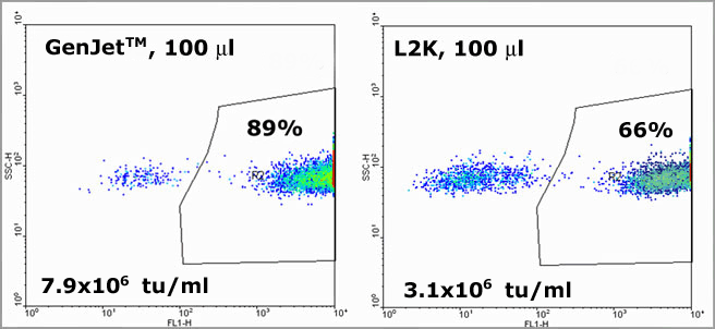
Exceptional high titers of lentivirus generated from 293F cells. Three DNAs co-transfected with GenJet™ In VitroDNA Transfection Reagent (left panel) in comparison of LipoFectamine 2000™ (L2K, right panel). 100 microliter virus supernatant from 293F cells were used to counter-infected 293F cells which was then passed FACS. The numbers at the upper right corner indicate the percentage of transduced cells. The titers of lentivirus generated with GenJet™ and L2K were quantified to be 7.9x10⁶ and 3.1x10⁶ tu/ml respectively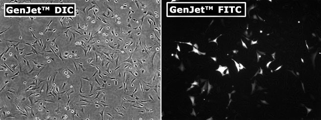
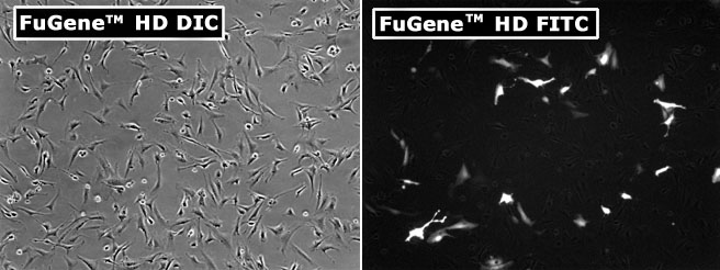
Primary Murine Skin Fibroblast transfected with pEGFP-C1 plasmid using GenJet™ In Vitro DNA Transfection Reagent (upper panel) and the most popular brand product FuGene™ HD (lower panel). The cells were visualized by Nikon Eclipse Fluorescence microscope with DIC phase imaging (left panel) and FITC imaging (right panel) 24 hours post-transfection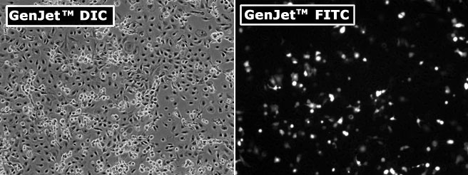
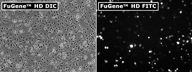
Primary Murine Keratinocytes transfected with pEGFP-C1 plasmid using GenJet™ In Vitro DNA Transfection Reagent (upper panel) and one of the most popular brand product FuGene™ HD (lower panel). The cells were visualized by Nikon Eclipse Fluorescence microscope with DIC phase imaging (left panel) and FITC imaging (right panel) 24 hours post-transfection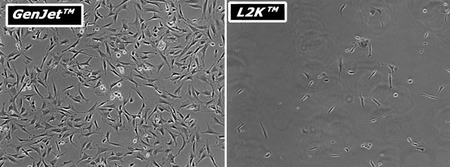
Comparison of cytotoxicity of GenJet™ DNA In Vitro Transfection Reagent with L2K™ on primary murine skin fibroblast. The primary murine fibroblast was incubated with the indicated transfection reagents/pEGFP-C1 (DNA) complexes above for 4 hours in serum-free DMEM High Glucose medium followed by replacement of complete serum-containing medium. The cells were visualized by Nikon Eclipse Fluorescence microscope with DIC phase imaging 24 hours post transfection